 Volume 109, Current Issue
 Vol 28, October 2019
 Vol 27, September 2019
 Vol 26, August 2019
 Vol 25, July 2019
 Vol 24, June 2019
 Vol 23, May 2019
 Vol 22, April 2019
 Vol 21, March 2019
 Vol 20, February 2019
 Vol 19, January 2019
 Vol 18, December 2018
 Vol 17, November 2018
 Vol 16, October 2018
 Vol 15, September 2018
 Vol 14, August 2018
 Vol 13, July 2018
 Vol 12, June 2018
 Vol 11, May 2018
 Vol 10, April 2018
 Vol 9, March 2018
 Vol 8, February 2018
 Vol 7, January 2018
 Vol 6 No 12, December 2017
 Vol 6 No 11, November 2017
 Vol 6 No 10, October 2017
 Vol 6 No 9, September 2017
 Vol 6 No 8, August 2017
 Vol 6 No 7, July 2017
 Vol 6 No 6, June 2017
 Vol 6 No 5, May 2017
 Vol 6 No 4, April 2017
 Vol 6 No 3, March 2017
 Vol 6 No 2, February 2017
 Vol 6 No 1, January 2017
 Vol 5 No 12, December 2016
 Vol 5 No 11, November 2016
 Vol 5 No 10, October 2016
 Vol 5 No 9, September 2016
 Vol 5 No 8, August 2016
 Vol 5 No 7, July 2016
 Vol 5 No 6, June 2016
 Vol 5 No 5, May 2016
 Vol 5 No 4, April 2016
 Vol 5 No 3, March 2016
 Vol 5 No 2, February 2016
 Vol 5 No 1, January 2016
 Vol 4 No 12, December 2015
 Vol 4 No 11, November 2015
 Vol 4 No 10, October 2015


Cover Story Current Issue

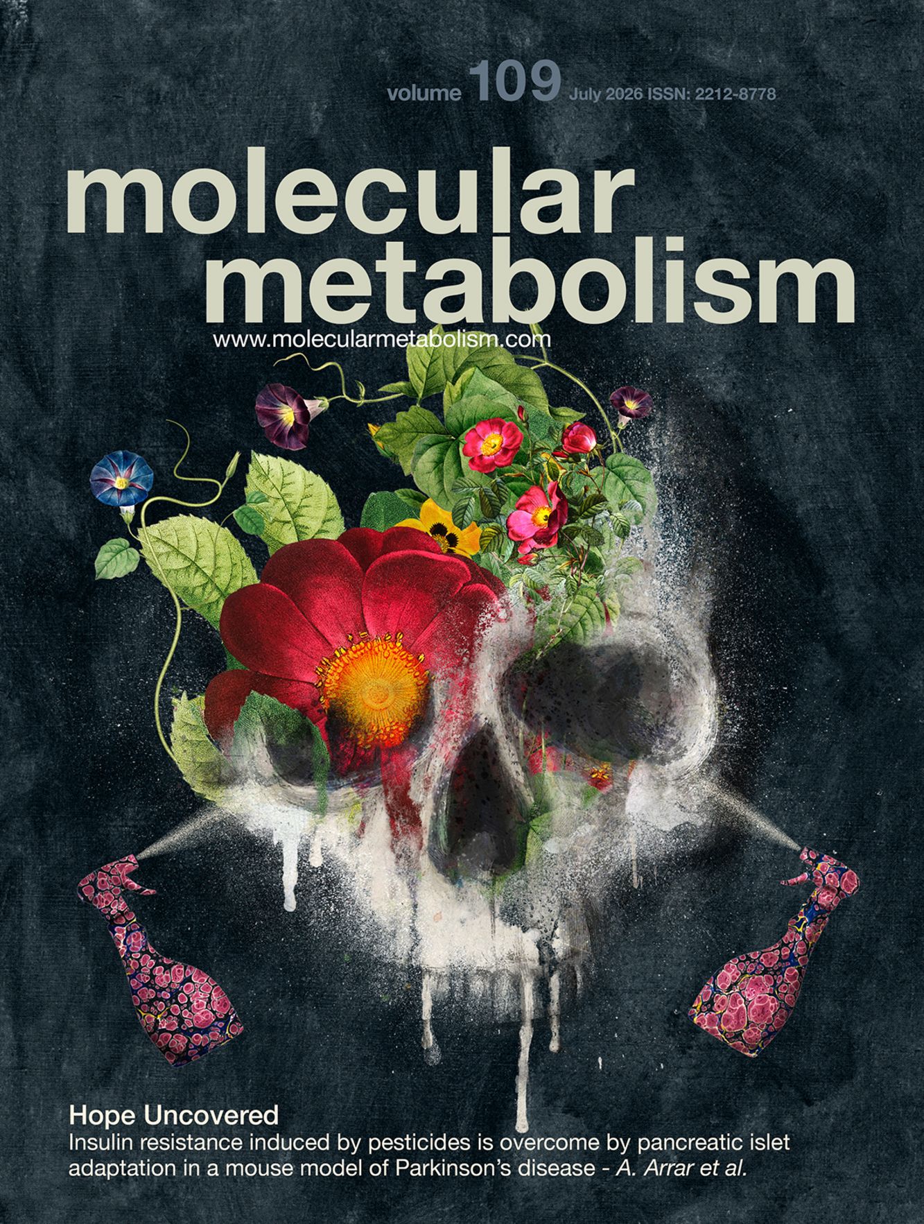
Epidemiological evidences provide proof of concept that certain pesticides are involved in metabolic disorders, but also in the pathophysiology of Parkinson's disease (PD). In addition, large prospective cohort studies reported that type 2 diabetes (T2D) and PD are epidemiologically associated, including an elevated risk of developing PD in patients with T2D.
Current Issue
miR-10a regulates cell death and inflammation in adipose tissue of male mice with diet-induced obesity
- Abstract
miR-10a regulates cell death and inflammation in adipose tissue of male mice with diet-induced obesity
Objective
Adipose tissue remodeling plays a critical role in obesity-induced metabolic dysfunction, but the underlying molecular mechanisms remain incompletely understood. This study investigates the role of miR-10a-5p in adipose tissue inflammation and metabolic dysfunction induced by a high-fat diet (HFD).
Methods
Male miR-10a knockout (KO) mice were fed a HFD to induce obesity for up to 16 weeks. RNA sequencing (RNA-seq) analysis was performed to profile mRNA expression and assess the effects of miR-10a-5p KO in gonadal white adipose tissue (gWAT). Additional analyses included immunoblotting, qPCR, histological examination, and validation of the miR-10a-5p target sequence using a dual-luciferase reporter assay.
Results
miR-10a-5p was highly expressed in gWAT but decreased after 8 weeks of HFD feeding. Over the 16-week HFD period, miR-10a KO mice exhibited greater weight gain and reduced energy expenditure compared to wild-type (WT) controls. gWAT of miR-10a KO mice on a HFD showed an increased population of proinflammatory macrophages, elevated inflammation, and increased cell death, characterized by upregulated apoptosis and necrosis markers. This was also associated with increased triglyceride accumulation in liver. Mechanistically, the proapoptotic gene Bcl2l11 was identified as a direct target of miR-10a-5p. Loss of miR-10a-5p led to BIM-mediated adipocyte death and inflammation, contributing to mitochondrial metabolic dysregulation, increased fibrosis marker expression, and the onset of inflammation in adipose tissue.
Conclusions
This study demonstrates the significant role of miR-10a-5p and its downstream target BIM in regulating adipocyte death during diet-induced obesity. This signaling pathway presents a potential therapeutic target for modulating obesity-induced inflammation and cell death in adipose tissue.
Articles in Press
miR-10a regulates cell death and inflammation in adipose tissue of male mice with diet-induced obesity
- Abstract
miR-10a regulates cell death and inflammation in adipose tissue of male mice with diet-induced obesity
Objective
Adipose tissue remodeling plays a critical role in obesity-induced metabolic dysfunction, but the underlying molecular mechanisms remain incompletely understood. This study investigates the role of miR-10a-5p in adipose tissue inflammation and metabolic dysfunction induced by a high-fat diet (HFD).
Methods
Male miR-10a knockout (KO) mice were fed a HFD to induce obesity for up to 16 weeks. RNA sequencing (RNA-seq) analysis was performed to profile mRNA expression and assess the effects of miR-10a-5p KO in gonadal white adipose tissue (gWAT). Additional analyses included immunoblotting, qPCR, histological examination, and validation of the miR-10a-5p target sequence using a dual-luciferase reporter assay.
Results
miR-10a-5p was highly expressed in gWAT but decreased after 8 weeks of HFD feeding. Over the 16-week HFD period, miR-10a KO mice exhibited greater weight gain and reduced energy expenditure compared to wild-type (WT) controls. gWAT of miR-10a KO mice on a HFD showed an increased population of proinflammatory macrophages, elevated inflammation, and increased cell death, characterized by upregulated apoptosis and necrosis markers. This was also associated with increased triglyceride accumulation in liver. Mechanistically, the proapoptotic gene Bcl2l11 was identified as a direct target of miR-10a-5p. Loss of miR-10a-5p led to BIM-mediated adipocyte death and inflammation, contributing to mitochondrial metabolic dysregulation, increased fibrosis marker expression, and the onset of inflammation in adipose tissue.
Conclusions
This study demonstrates the significant role of miR-10a-5p and its downstream target BIM in regulating adipocyte death during diet-induced obesity. This signaling pathway presents a potential therapeutic target for modulating obesity-induced inflammation and cell death in adipose tissue.
Registration & Abstract Submission are open!
13th
Helmholtz Diabetes Conference
Munich, 21-23. Sep 2026
2024 impact factor: 6.6
You are what you eat
Here is a video of Vimeo. When the iframes is activated, a connection to Vimeo is established and, if necessary, cookies from Vimeo are also used. For further information on cookies policy click here.